Designing a Labeled Coloring Sheet

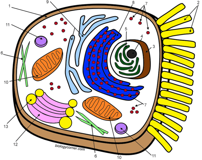
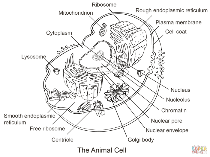
Animal cell labeled coloring sheet – Creating a visually engaging and informative animal cell coloring sheet requires careful consideration of layout and clarity. The goal is to present the key organelles in a way that is both aesthetically pleasing and easy for students to understand and label. A well-designed sheet will aid in memorization and comprehension of animal cell structure.This section details the design process for such a coloring sheet, focusing on the arrangement of organelles and the creation of clear labels.
We will use a simple diagram as a basis for the coloring sheet design, ensuring that the organelles are clearly distinguishable and appropriately labeled.
Animal Cell Diagram and Organelle Placement
The animal cell diagram should be simple yet comprehensive. It should include the major organelles: the nucleus, cytoplasm, cell membrane, mitochondria, ribosomes, endoplasmic reticulum (both rough and smooth), Golgi apparatus, lysosomes, and vacuoles. The size and placement of these organelles should reflect their relative proportions within a typical animal cell, though perfect scale isn’t necessary for a coloring sheet.
The nucleus should be centrally located and relatively large, while mitochondria should be depicted as numerous small bean-shaped structures scattered throughout the cytoplasm. The endoplasmic reticulum can be shown as a network of interconnected membranes, with the rough ER appearing studded with ribosomes. The Golgi apparatus can be represented as a stack of flattened sacs. Lysosomes and vacuoles can be shown as smaller, round structures.
The cell membrane should form a clear boundary around the entire cell.
Coloring Sheet Design and Organelle Labels
The coloring sheet will utilize a table format to organize the organelles and their descriptions. This approach provides a structured way to present information, making it easy for students to locate and label each component. The table will consist of four columns for optimal readability and visual appeal on various screen sizes. Each row will represent a different organelle, including its name, a short description of its function, and a suggested color for coloring.
| Organelle | Description | Suggested Color | Location on Diagram |
|---|---|---|---|
| Nucleus | Contains the cell’s genetic material (DNA). | Light purple | Center of the cell |
| Cytoplasm | The jelly-like substance filling the cell. | Light yellow | Fills the space inside the cell membrane |
| Cell Membrane | The outer boundary of the cell, regulating what enters and exits. | Dark blue | Outermost layer of the cell |
| Mitochondria | Powerhouses of the cell, producing energy (ATP). | Bright red | Scattered throughout the cytoplasm |
| Ribosomes | Sites of protein synthesis. | Dark green | Attached to the rough ER or free in the cytoplasm |
| Endoplasmic Reticulum (Rough) | Network of membranes studded with ribosomes; involved in protein synthesis and transport. | Light green | Network extending from the nucleus |
| Endoplasmic Reticulum (Smooth) | Network of membranes involved in lipid synthesis and detoxification. | Pale green | Network extending from the nucleus |
| Golgi Apparatus | Processes and packages proteins and lipids. | Orange | Near the nucleus |
| Lysosomes | Contain enzymes that break down waste materials. | Brown | Scattered throughout the cytoplasm |
| Vacuoles | Storage sacs for water, nutrients, and waste. | Light blue | Scattered throughout the cytoplasm |
Coloring Sheet Content and Detail
This section details the function of each labeled organelle within an animal cell, providing interesting facts and highlighting key differences between animal and plant cells. Understanding these organelles is crucial to grasping the complexities of cellular life and the fundamental differences between these two major cell types.The following descriptions provide a comprehensive overview of the major organelles typically included in an animal cell coloring sheet, focusing on their roles and unique characteristics.
Understanding animal cell structures can be fun and engaging, especially for younger learners. A well-labeled animal cell coloring sheet provides a great visual aid for grasping the functions of organelles like the nucleus and mitochondria. For a slightly different, yet related, creative activity, you might also consider checking out some animal bunny coloring pages which offer a change of pace while still focusing on the animal kingdom.
Returning to the cellular level, remember that detailed coloring sheets are invaluable tools for reinforcing learning about animal cells.
We will explore their individual functions and compare them to their counterparts (or lack thereof) in plant cells.
Cell Membrane
The cell membrane is the outer boundary of the cell, a selectively permeable barrier that controls what enters and exits. It’s composed of a phospholipid bilayer with embedded proteins. Think of it as a sophisticated gatekeeper, allowing essential nutrients in and waste products out. This dynamic structure is constantly changing and adapting to the cell’s needs. Plant cells also have a cell membrane, but they possess an additional outer layer, the cell wall, providing structural support.
Cytoplasm
The cytoplasm is the jelly-like substance filling the cell, excluding the nucleus. It’s a dynamic environment where many cellular processes occur. Organelles are suspended within the cytoplasm, and it plays a crucial role in cell signaling and transport. Both animal and plant cells contain cytoplasm, though the composition might differ slightly.
Nucleus
The nucleus is the control center of the cell, containing the cell’s genetic material (DNA). It’s surrounded by a double membrane called the nuclear envelope, which regulates the movement of molecules in and out. The nucleus directs the cell’s activities by controlling gene expression. Both animal and plant cells have a nucleus.
Ribosomes
Ribosomes are the protein factories of the cell. They are responsible for translating the genetic code from DNA into proteins, the building blocks of life. These tiny organelles can be found free-floating in the cytoplasm or attached to the endoplasmic reticulum. Both animal and plant cells contain ribosomes.
Endoplasmic Reticulum (ER)
The endoplasmic reticulum is a network of interconnected membranes extending throughout the cytoplasm. There are two types: rough ER (studded with ribosomes) and smooth ER (lacking ribosomes). Rough ER is involved in protein synthesis and modification, while smooth ER plays a role in lipid synthesis and detoxification. Both animal and plant cells possess an endoplasmic reticulum.
Golgi Apparatus (Golgi Body)
The Golgi apparatus is the cell’s packaging and processing center. It modifies, sorts, and packages proteins and lipids received from the ER, preparing them for transport to other parts of the cell or secretion outside the cell. Both animal and plant cells utilize a Golgi apparatus.
Mitochondria, Animal cell labeled coloring sheet
Mitochondria are the powerhouses of the cell, generating energy in the form of ATP (adenosine triphosphate) through cellular respiration. They have their own DNA and ribosomes, suggesting an endosymbiotic origin. Both animal and plant cells contain mitochondria, although plant cells also generate energy through photosynthesis.
Lysosomes
Lysosomes are the cell’s recycling centers, containing digestive enzymes that break down waste products, cellular debris, and foreign invaders. They maintain cellular health by removing unwanted materials. While present in animal cells, plant cells typically utilize vacuoles for similar functions.
Vacuoles
Vacuoles are membrane-bound sacs used for storage. In animal cells, they are generally small and numerous, involved in various functions, including waste storage. Plant cells, however, typically possess a large central vacuole that occupies a significant portion of the cell’s volume, contributing to turgor pressure and storage of water and nutrients. This is a key difference between animal and plant cells.
Variations and Extensions of the Coloring Sheet Design

This section explores several ways to adapt the basic animal cell coloring sheet design, catering to different age groups and learning styles. Expanding upon the core design allows for increased engagement and a deeper understanding of cell biology. We will examine more complex, simplified, and interactive versions of the coloring sheet.
A More Complex Animal Cell Coloring Sheet
This version incorporates additional organelles, providing a more comprehensive view of the cell’s structure and function. The inclusion of these additional structures offers a more challenging and rewarding activity for older students or those with a greater interest in biology. The added organelles could include the endoplasmic reticulum (both rough and smooth), Golgi apparatus, lysosomes, peroxisomes, and centrioles.
The detailed illustration would show the spatial relationships between these organelles and those already present in the basic design. For example, the rough endoplasmic reticulum could be depicted studded with ribosomes, clearly indicating its role in protein synthesis. Similarly, the Golgi apparatus could be illustrated as a series of flattened sacs, highlighting its function in modifying and packaging proteins.
Clear labeling of each organelle and its function is crucial for effective learning.
A Simplified Animal Cell Coloring Sheet for Younger Learners
For younger learners, a simplified version focusing on only the major organelles is beneficial. This reduces cognitive overload and allows them to grasp the fundamental concepts more easily. This simplified version would include only the nucleus, cytoplasm, cell membrane, mitochondria, and ribosomes. The illustrations would be larger, bolder, and less detailed, making them easier for young children to color and identify.
The labels could be larger and use simpler language, avoiding complex terminology. For instance, instead of “mitochondria,” the label could simply read “powerhouse of the cell.” The overall design should be more visually appealing, potentially incorporating bright colors and playful elements.
An Interactive Animal Cell Coloring Sheet with Fill-in-the-Blank Labels
This interactive version incorporates a fill-in-the-blank activity, enhancing engagement and promoting active learning. The coloring sheet would include the illustration of an animal cell with blank spaces next to each organelle. Students would then need to fill in the correct name of the organelle based on their knowledge or by using a provided word bank. This activity encourages students to actively recall and apply their understanding of cell structures.
This version could also include short descriptions or functions of each organelle in a separate section for students to reference while completing the fill-in-the-blank activity. A possible extension would be to add a section where students could draw and label additional organelles not included in the main illustration, encouraging further research and exploration.
Commonly Asked Questions: Animal Cell Labeled Coloring Sheet
Where can I find printable versions of this coloring sheet?
Printable versions can be easily created from digital copies using a standard printer. Many online resources may also offer printable versions.
What age group is this coloring sheet suitable for?
The complexity can be adjusted. A simplified version is suitable for younger learners (elementary school), while a more detailed version is appropriate for older students (middle and high school).
Can this coloring sheet be used for homeschooling?
Absolutely! It’s a perfect supplement for homeschooling biology curricula, offering a hands-on, visual learning experience.
Are there any online interactive versions available?
While not directly included, the design could be adapted for interactive online platforms, allowing for digital labeling and quizzes.


