Cell Organelles and Their Coloration: Animal Cell Coloring Questions And Answers

Animal cell coloring questions and answers – Understanding the appearance of different cell organelles under a microscope is crucial for accurate cell identification and analysis. The staining techniques used significantly impact the visual characteristics of these organelles, allowing for their differentiation and study. This section will detail the typical appearance of several key animal cell organelles following common staining procedures.
Nucleus Appearance
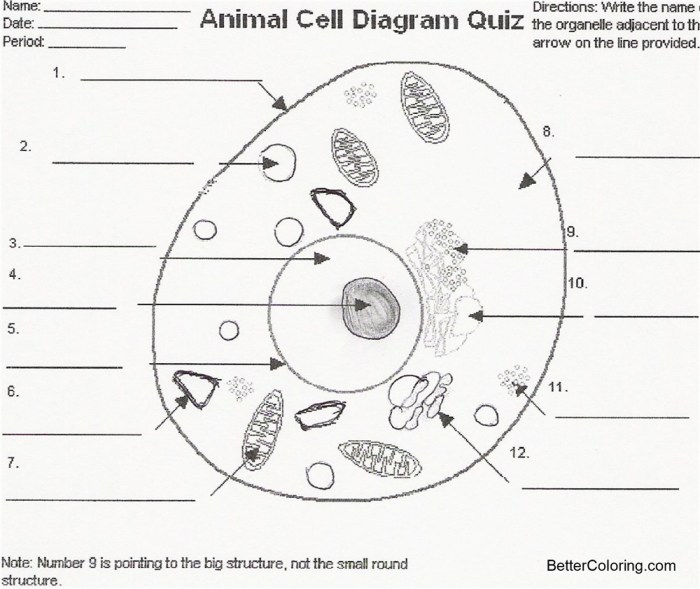
In a stained animal cell, the nucleus typically appears as a large, round or oval structure. The chromatin, a complex of DNA and proteins, stains intensely, usually a dark purple or deep blue, depending on the stain used (e.g., hematoxylin). This dark coloration contrasts sharply with the lighter surrounding cytoplasm. The nucleolus, a smaller, round structure within the nucleus, may also be visible, often appearing as a slightly lighter shade within the intensely stained chromatin.
The nuclear envelope, while often less intensely stained than the chromatin, may be visible as a faint boundary surrounding the nucleus.
Cytoplasm Appearance
The cytoplasm, the gel-like substance filling the cell, generally appears as a light pink or pale blue in commonly used stains like eosin or methylene blue. Its texture is usually granular, reflecting the presence of various organelles and cellular components. The exact shade and granularity will depend on the type and concentration of the stain used, as well as the cell type itself.
Differences in staining intensity within the cytoplasm can sometimes indicate regions of higher or lower organelle density.
Endoplasmic Reticulum Structure Revealed by Staining
Different staining techniques can reveal the intricate structure of the endoplasmic reticulum (ER). For instance, electron microscopy, though not a staining technique in the traditional sense, provides the highest resolution images, showcasing the ER as a network of interconnected membranes. Specific stains used in light microscopy, while not providing the same level of detail, can differentiate between rough ER (studded with ribosomes) and smooth ER.
Rough ER often appears slightly darker than the surrounding cytoplasm due to the ribosomes bound to its surface, while smooth ER may appear as a more diffuse, lighter network.
Golgi Apparatus Characteristics
The Golgi apparatus, typically located near the nucleus, usually stains lighter than the nucleus but darker than the surrounding cytoplasm. Its characteristic layered structure, consisting of flattened sacs (cisternae), may be visible under higher magnification as a stacked arrangement of these lightly stained membranes. The precise appearance depends heavily on the staining method and the cell’s physiological state, with some stains potentially highlighting the Golgi more intensely than others.
Understanding animal cell coloring questions and answers can be a fun and educational experience, especially when you consider the intricate details of each organelle. This knowledge can be further enhanced by exploring the larger context of animal anatomy; for instance, you might find helpful visual aids in resources like these animal body parts coloring pages , which provide a broader perspective on how these cells contribute to the overall organism.
Returning to the cellular level, remember that accurate coloring helps solidify your comprehension of animal cell structures and their functions.
Diagram of Stained Animal Cell Organelles
Imagine a circular cell. The nucleus, a large dark purple circle, sits near the center. Surrounding it is a pale pink cytoplasm speckled with small, darker purple dots representing ribosomes. A network of slightly darker pink strands represents the endoplasmic reticulum, with some regions appearing slightly darker, indicating rough ER. Near the nucleus, a stack of pale, slightly darker pink flattened sacs represents the Golgi apparatus.
Other organelles, such as mitochondria (represented as small, dark reddish-purple oval shapes) and lysosomes (represented as tiny dark purple dots) are scattered throughout the cytoplasm. The cell membrane is represented by a thin, dark line forming the outer boundary of the cell. The relative sizes and staining intensities of these organelles are approximate representations and can vary depending on the cell type and staining method.
Cell Processes and Visual Representation
Understanding cell processes is crucial for comprehending the intricacies of life. Visual representations aid in this understanding, allowing us to see the dynamic nature of cellular activities. This section explores key cellular processes and how they can be depicted visually.
Mitosis and Chromosome Appearance
Mitosis, the process of cell division, is characterized by distinct stages, each with a unique chromosomal arrangement. Observing these stages visually helps clarify the process of cell replication.
- Prophase: Chromosomes condense and become visible as distinct X-shaped structures. The nuclear envelope begins to break down.
- Metaphase: Chromosomes align along the metaphase plate, an imaginary line in the center of the cell. This alignment ensures equal distribution of genetic material to daughter cells.
- Anaphase: Sister chromatids separate and move towards opposite poles of the cell, pulled by spindle fibers. The cell elongates.
- Telophase: Chromosomes reach the poles, decondense, and become less visible. The nuclear envelope reforms around each set of chromosomes. Cytokinesis, the division of the cytoplasm, begins.
- Cytokinesis: The cell divides completely, resulting in two genetically identical daughter cells.
Visual Representation of Active Transport
Active transport, the movement of molecules against their concentration gradient, requires energy. In a cell model, this could be visually represented by depicting molecules moving from a region of low concentration to a region of high concentration. Arrows indicating the direction of movement and a distinct color or shading for the energy source (e.g., ATP) could further enhance the representation.
For instance, a protein pump actively transporting sodium ions out of the cell could be shown with sodium ions moving against their concentration gradient, with the pump being clearly labelled and ATP molecules depicted nearby.
Distinguishing Visual Cues of Different Animal Cells
Different animal cells have distinct shapes and structures reflecting their specialized functions.Muscle cells, for example, are long and cylindrical, often exhibiting striations (striped appearance) due to the organized arrangement of contractile proteins. Nerve cells, or neurons, are characterized by their long, branching extensions (axons and dendrites) that facilitate the transmission of nerve impulses. Epithelial cells, forming linings, often have a tightly packed arrangement and distinct shapes depending on their location.
This visual diversity highlights the cellular specialization within an organism.
Comparison of Healthy and Unhealthy Animal Cells
Microscopic examination reveals significant differences between healthy and unhealthy animal cells.
| Feature | Healthy Cell | Unhealthy Cell | Visual Cue |
|---|---|---|---|
| Cell Membrane | Intact, smooth | Damaged, irregular | Smooth, defined Artikel vs. blebbing or disrupted edges |
| Cytoplasm | Homogeneous, clear | Granular, vacuolated | Uniform texture vs. presence of dark spots or vacuoles |
| Nucleus | Well-defined, round | Shrunken, irregular, or fragmented | Clear, centrally located nucleus vs. shrunken or damaged nucleus |
| Organelles | Normally sized and shaped | Swollen, damaged, or absent | Presence and proper morphology of organelles vs. abnormal size, shape, or absence |
Visual Representation of Osmosis and Diffusion
Imagine a cell immersed in a solution. For diffusion, depict small solute molecules moving randomly across the cell membrane from an area of high concentration to an area of low concentration, represented by arrows indicating the direction of movement. For osmosis, show water molecules moving across the selectively permeable membrane from an area of high water concentration (low solute concentration) to an area of low water concentration (high solute concentration).
The direction of water movement will depend on the concentration gradient across the membrane. Different colors or sizes for water and solute molecules would enhance clarity. The cell might swell (hypotonic solution) or shrink (hypertonic solution) depending on the surrounding solution’s concentration, visually demonstrating the effect of osmosis.
Microscopy and Staining Techniques

Microscopy and staining techniques are fundamental to visualizing the intricate structures within animal cells. Understanding these techniques allows researchers and students to identify and study various organelles, revealing the complex processes occurring within these microscopic units of life. The choice of technique depends heavily on the specific structures of interest and the level of detail required.
Importance of Staining Techniques in Visualizing Cell Structures
Staining techniques are crucial because animal cells are largely transparent. Without staining, many organelles would be invisible under a microscope. Different stains bind to specific cellular components, highlighting their structures and enabling their identification. For example, hematoxylin stains the nucleus a dark purple, while eosin stains the cytoplasm pink, providing a clear contrast between these two major cellular regions.
The selective nature of these stains allows for the differentiation and identification of various cellular structures, leading to a deeper understanding of cell biology.
Procedure for Preparing an Animal Cell Slide for Microscopic Observation, Animal cell coloring questions and answers
Preparing a slide for microscopic observation involves several steps. First, a small sample of animal tissue (e.g., cheek cells) is obtained. This sample is then smeared thinly onto a clean glass slide to create a single layer of cells. Next, the slide is allowed to air dry completely. Following this, a fixative (such as methanol) is applied to preserve the cell structure and prevent degradation.
After fixation, the slide is stained using the appropriate technique, rinsed gently, and then mounted with a coverslip using a mounting medium. Finally, the slide is ready for observation under a microscope.
Comparison of Light and Electron Microscopes
Light microscopes use visible light to illuminate the specimen, offering a relatively simple and inexpensive method for observing cells. However, their resolution is limited, restricting the visualization of very small organelles. Electron microscopes, on the other hand, use a beam of electrons to illuminate the specimen, achieving significantly higher resolution. This allows for the visualization of much smaller structures, such as ribosomes and the internal structures of organelles.
However, electron microscopy requires specialized equipment, extensive preparation techniques, and is considerably more expensive than light microscopy. Light microscopy provides a good overview of cell structure, while electron microscopy provides detailed ultrastructural information.
Impact of Staining Techniques on the Color and Visibility of Specific Organelles
Different stains target specific cellular components, affecting their color and visibility. For instance, hematoxylin stains negatively charged molecules like DNA in the nucleus, making it intensely dark purple. Eosin, on the other hand, stains positively charged components like proteins in the cytoplasm, resulting in a pink or reddish hue. Specific stains, such as the Golgi stain, are used to visualize the Golgi apparatus, which would otherwise be difficult to distinguish.
The choice of stain directly impacts the clarity and contrast of the observed organelles, allowing researchers to focus on specific structures of interest.
Creating a Labeled Diagram of an Animal Cell from Microscopic Observation
To create a labeled diagram, begin by carefully observing the prepared slide under the microscope. Identify the different organelles and note their shapes, sizes, and locations within the cell. Next, draw a detailed representation of the cell, accurately reflecting the observed structures. Finally, label each identified organelle using clear and concise labels, referencing the stain used and the observed characteristics of each structure.
The diagram should accurately represent the cell’s structure as observed under the microscope, providing a visual record of the microscopic observation.
Frequently Asked Questions
What is the best stain to visualize the cell membrane?
Various stains can highlight the cell membrane, depending on the desired level of detail. Common choices include methylene blue and crystal violet, which generally stain the membrane a dark blue or purple.
How do I prepare a good animal cell slide for microscopy?
Proper slide preparation is crucial. This typically involves obtaining a sample (e.g., cheek cells), spreading it thinly on a slide, staining it with an appropriate dye, and then adding a coverslip to protect the sample.
What are some limitations of using light microscopy to observe animal cells?
Light microscopy has limitations in resolution; very small organelles might not be clearly visible. Electron microscopy offers much higher resolution but is more complex and expensive.
Why is understanding animal cell structure important?
A thorough understanding of animal cell structure is fundamental to comprehending biological processes, disease mechanisms, and developing effective treatments. It underpins various fields like medicine, biotechnology, and agriculture.


