Key Organelles and Their Functions: Animal Cell Coloring Diagram Key
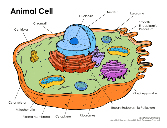
Animal cell coloring diagram key – Animal cells are complex structures containing various organelles, each with specialized roles crucial for the cell’s survival and function. Understanding these organelles and their interactions is fundamental to comprehending cellular biology. This section will detail the structure and function of several key organelles within the animal cell.
Mitochondria: The Powerhouses of the Cell
Mitochondria are double-membraned organelles often described as the “powerhouses” of the cell. The outer membrane is smooth, while the inner membrane is highly folded into cristae, significantly increasing its surface area. This intricate structure is essential for the mitochondrion’s primary function: cellular respiration. Through a series of metabolic processes, mitochondria break down glucose and other fuel molecules to generate adenosine triphosphate (ATP), the cell’s primary energy currency.
The cristae provide ample space for the protein complexes involved in the electron transport chain, a crucial step in ATP production. Mitochondria also play a role in other cellular processes, including calcium storage and apoptosis (programmed cell death). Their own independent DNA further highlights their unique role within the cell.
Endoplasmic Reticulum: A Network of Membranes
The endoplasmic reticulum (ER) is an extensive network of interconnected membranes extending throughout the cytoplasm. There are two distinct types: rough ER and smooth ER, each with unique characteristics and functions. The rough ER, studded with ribosomes, is involved in protein synthesis and modification. Ribosomes on the rough ER synthesize proteins destined for secretion, insertion into membranes, or transport to other organelles.
The smooth ER, lacking ribosomes, plays a key role in lipid synthesis, detoxification of harmful substances, and calcium storage. Its smooth surface reflects its different functions compared to the ribosome-studded rough ER.
Golgi Apparatus and Lysosomes: Processing and Recycling Centers
The Golgi apparatus, also known as the Golgi complex, is a stack of flattened, membrane-bound sacs called cisternae. It receives proteins and lipids synthesized by the ER and further processes, modifies, and sorts them for transport to their final destinations within or outside the cell. This crucial sorting and packaging function ensures that molecules reach their correct locations within the complex cellular environment.
Lysosomes, on the other hand, are membrane-bound organelles containing hydrolytic enzymes. They act as the cell’s recycling centers, breaking down waste products, cellular debris, and ingested materials. This degradation process is essential for maintaining cellular homeostasis and preventing the accumulation of harmful substances.
Ribosomes and Vacuoles: A Comparison
The following points compare and contrast the functions of ribosomes and vacuoles:
- Ribosomes: Synthesize proteins according to the instructions encoded in messenger RNA (mRNA). They are found free in the cytoplasm or bound to the rough ER. Their primary function is protein synthesis.
- Vacuoles: Membrane-bound sacs that store various substances, including water, nutrients, and waste products. In animal cells, vacuoles are generally smaller and more numerous than in plant cells. Their primary function is storage and maintenance of turgor pressure (though less significant in animal cells than plants).
Coloring and Representation of Cell Components
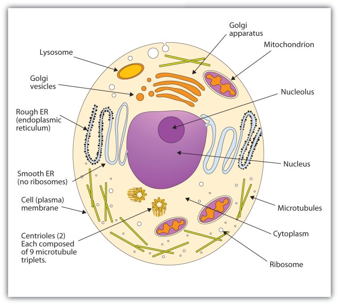
Creating a visually informative and accurate animal cell diagram requires a thoughtful approach to color-coding and the representation of organelle size and membrane structure. A well-designed diagram enhances understanding by clearly distinguishing different cellular components and their relative importance.Color selection plays a crucial role in the clarity and memorability of the diagram. Careful consideration of color theory and the symbolic associations of different colors can significantly improve the overall effectiveness of the visualization.
Color-Coding Scheme for Animal Cell Organelles
The following color scheme provides a unique color for each major animal cell organelle. The rationale behind each choice considers both visual contrast and potential mnemonic associations.
- Nucleus: Dark Purple. The deep color represents the nucleus’s central role and importance as the cell’s control center.
- Nucleolus: Lighter Purple. A slightly lighter shade distinguishes the nucleolus within the nucleus, indicating its function as a ribosome factory.
- Rough Endoplasmic Reticulum (RER): Dark Blue. The darker shade emphasizes the RER’s role in protein synthesis, reflecting its studded appearance with ribosomes.
- Smooth Endoplasmic Reticulum (SER): Light Blue. The lighter shade contrasts with the RER, signifying its different functions in lipid synthesis and detoxification.
- Ribosomes: Dark Gray. The neutral color avoids overshadowing other organelles while representing their ubiquitous presence.
- Golgi Apparatus: Light Green. The light green color represents the Golgi’s role in packaging and processing proteins and lipids, associating it with growth and renewal.
- Mitochondria: Red. The vibrant color represents the mitochondria’s energy-producing function, symbolizing power and activity.
- Lysosomes: Orange. The bright color suggests the lysosomes’ role in waste breakdown and digestion.
- Peroxisomes: Yellow. Yellow suggests the peroxisome’s role in breaking down fatty acids and other molecules, generating hydrogen peroxide as a byproduct.
- Cytoskeleton: Light Brown. The muted color represents the structural support of the cell, subtly highlighting its presence without overwhelming other organelles.
- Plasma Membrane: Black. The strong Artikel provides clear definition and contrast to the internal structures.
- Cytoplasm: Pale Yellow. The pale background color provides a neutral space for the organelles, allowing them to stand out clearly.
Representing Relative Sizes of Organelles
Accurately depicting the relative sizes of organelles is crucial for creating a realistic and informative cell diagram. This can be achieved through careful scaling based on actual size ratios. For example, the nucleus is generally the largest organelle, followed by the mitochondria, then the other organelles in decreasing order of size. While exact scaling to microscopic dimensions isn’t necessary, maintaining relative proportions is essential.
A simple method would be to establish a scale where the nucleus, being the largest, is represented at a certain size, and then scale other organelles proportionally smaller.
Depicting the Membrane Structure
The plasma membrane and other membranes within the cell (e.g., those surrounding the nucleus and organelles) should be represented as a double-layered structure. This can be depicted as two parallel lines, separated by a small gap, to represent the phospholipid bilayer. The proteins embedded within the membrane can be shown as small, differently colored shapes interspersed within the bilayer.
Understanding an animal cell coloring diagram key is crucial for visualizing cellular structures. This detailed knowledge contrasts sharply with the often simplified representations found in activities like the animal captivity coloring puzzle , which focuses on animal forms rather than their microscopic components. Returning to the cellular level, the key helps accurately identify organelles and their functions within the animal cell.
This representation accurately reflects the fluid mosaic model of membrane structure, highlighting its dynamic and complex nature. The space between the two lines of the bilayer should be noticeably thinner than the lines themselves, accurately reflecting the membrane’s overall thinness relative to the size of the organelles.
Creating a Diagram with Key
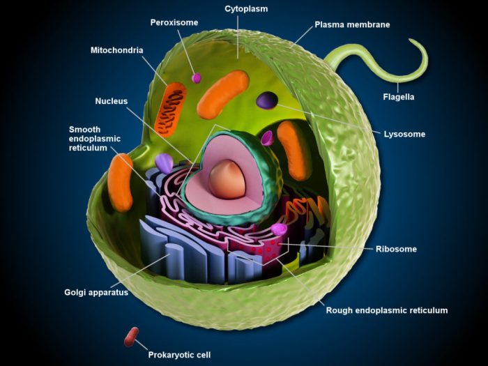
Creating a visually engaging and informative animal cell diagram requires careful organization of information and a well-designed key. This section details the process of constructing a table to organize your data, building a legend using HTML, and providing tips for creating a clear and appealing diagram.
To effectively represent the various organelles within an animal cell, a structured approach is necessary. This involves compiling data about each organelle, assigning a color for visual representation, and then using this information to create a key that viewers can easily reference.
Organelle Data Table, Animal cell coloring diagram key
Before creating the diagram itself, it’s helpful to organize your information into a table. This table will serve as the foundation for both your diagram and its accompanying key. This structured approach ensures consistency and accuracy in your representation.
| Organelle Name | Function | Color |
|---|---|---|
| Nucleus | Contains genetic material (DNA); controls cell activities. | Dark Purple |
| Cell Membrane | Regulates what enters and leaves the cell; protects the cell. | Light Blue |
| Cytoplasm | Gel-like substance filling the cell; supports organelles. | Light Green |
| Mitochondria | Powerhouse of the cell; produces energy (ATP). | Red |
| Ribosomes | Synthesize proteins. | Dark Brown |
| Endoplasmic Reticulum (ER) | Network of membranes; transports materials within the cell. (Rough ER has ribosomes attached.) | Light Orange (Rough ER: slightly darker orange) |
| Golgi Apparatus | Processes and packages proteins. | Yellow |
| Lysosomes | Break down waste materials and cellular debris. | Dark Green |
| Centrioles | Involved in cell division. | Pink |
Creating a Diagram Key using HTML
The key, or legend, is crucial for understanding your diagram. A well-formatted HTML table provides a clear and easily accessible way to present this information. Using responsive design principles ensures readability across different screen sizes.
Below is an example of how to create a key using HTML table tags. Remember to replace the example colors with the colors you chose in your data table.
| Organelle Name | Color |
|---|---|
| Nucleus | |
| Cell Membrane | |
| Cytoplasm | |
| Mitochondria | |
| Ribosomes | |
| Endoplasmic Reticulum (ER) | |
| Golgi Apparatus | |
| Lysosomes | |
| Centrioles |
Tips for Creating a Visually Appealing Diagram
To ensure your diagram is both informative and aesthetically pleasing, consider these tips:
Use clear and concise labels. Avoid overcrowding the diagram with excessive text. Maintain a consistent scale for the organelles to accurately reflect their relative sizes. Employ a visually appealing color scheme that enhances clarity and avoids clashing colors. Consider using different textures or patterns to further differentiate organelles if necessary.
A well-organized layout with sufficient spacing between organelles will enhance readability. Finally, ensure that the diagram is large enough to be easily viewed and understood, yet not excessively large.
Frequently Asked Questions
What are some common mistakes to avoid when creating an animal cell diagram?
Common mistakes include inaccurate representation of organelle sizes, inconsistent coloring, and unclear labeling. Ensure organelles are proportionally sized, colors are consistent with your key, and labels are clear and concise.
Why is color-coding important in a cell diagram?
Color-coding enhances clarity and visual appeal, making it easier to identify and distinguish different organelles. A well-chosen color scheme improves understanding and memorization.
Are there any online resources available to help create animal cell diagrams?
Yes, many online resources offer templates, tutorials, and even interactive tools for creating cell diagrams. A simple web search for “animal cell diagram maker” will yield many results.

