Interpreting Colored Animal Cell Diagrams

Animal cell coloring answers front – Colored diagrams of animal cells are invaluable tools for understanding cell structure and function. By assigning different colors to various organelles, these diagrams visually represent the complexity of cellular components and their interactions. Effective interpretation requires understanding how color choices relate to organelle function and recognizing the limitations of this representation method.
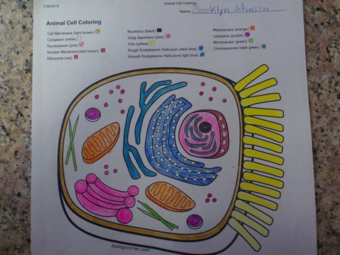
The effective use of color in a cell diagram simplifies the visualization of intricate cellular structures and processes. For instance, the nucleus, often depicted in a darker shade like purple or dark blue, immediately stands out as the control center containing the cell’s genetic material. Similarly, the bright green often used for chloroplasts in plant cell diagrams highlights their role in photosynthesis; however, this would be absent in an animal cell diagram.
Instead, mitochondria, crucial for energy production, might be colored bright red or orange to emphasize their metabolic activity.
Key Organelle Identification and Color-Function Correlation
A well-designed colored animal cell diagram will clearly identify major organelles such as the nucleus (often dark purple or blue), ribosomes (small dots, perhaps gray or dark brown), the endoplasmic reticulum (ER) (a network, possibly light blue for rough ER and light pink for smooth ER), the Golgi apparatus (stacked structures, maybe light green or yellow), mitochondria (often red or orange), lysosomes (small, possibly dark red or purple), and the cell membrane (outer boundary, usually a thin line of dark brown or black).
The color choices reflect the organelle’s function; for example, the dark color of the nucleus emphasizes its importance as the information center. The different shades of the ER represent its varied roles in protein synthesis and lipid metabolism.
Limitations of Color Representation in Cellular Processes
While color coding is helpful, it presents limitations. Complex processes like protein synthesis or cellular respiration, involving multiple organelles, are simplified in a static image. The diagram cannot fully represent the dynamic nature of these processes, the constant movement of molecules, or the temporal changes within the cell. Further, color interpretation is subjective; different diagrams may use different color schemes, leading to potential confusion.
The three-dimensional nature of the cell is also reduced to a two-dimensional representation, obscuring the spatial relationships between organelles.
Understanding Organelle Relationships Using Colored Diagrams
Colored diagrams help visualize the relationships between organelles. For example, the proximity of ribosomes to the rough endoplasmic reticulum clearly shows the role of the ER in protein synthesis, where ribosomes produce proteins that are then transported through the ER. The connection between the ER and the Golgi apparatus illustrates the pathway of protein modification and transport. The distribution of mitochondria throughout the cytoplasm highlights their role in providing energy to all parts of the cell.
Finding the answers for your animal cell coloring sheet might seem challenging at first, but remember that understanding the structures is key. If you need a break from the complexities of cell biology, consider checking out some fun coloring pages, such as these animal alphabet j coloring pages printable , for a refreshing change of pace. Returning to your animal cell diagram, remember to carefully label each organelle before moving on to the next coloring activity.
The close association of lysosomes with other organelles suggests their involvement in waste removal and recycling.
Color Highlighting of Specific Cellular Processes or Structures
Color can highlight specific structures or processes. For example, a brighter shade of red could be used to emphasize a particular area of high metabolic activity within the mitochondria. A different color could distinguish between different types of vesicles or highlight the cytoskeleton’s structural role. In diagrams depicting cell division, different colors might represent chromosomes at various stages of mitosis or meiosis.
A cell undergoing apoptosis (programmed cell death) could be visually distinguished through the use of a specific color palette to emphasize the changes in organelle morphology and function.
Creating Educational Resources about Animal Cells: Animal Cell Coloring Answers Front

Developing effective educational resources is crucial for fostering a deep understanding of animal cell biology. By utilizing diverse teaching methods, we can cater to various learning styles and enhance knowledge retention. The following Artikels the creation of several resources aimed at improving student comprehension of animal cell structure and function.
Animal Cell Worksheet Design
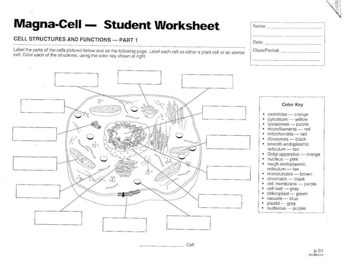
This worksheet will feature a large, blank diagram of an animal cell. Students will be required to color-code different organelles (e.g., nucleus – purple, mitochondria – red, Golgi apparatus – blue, etc.), using a provided key or legend. Following the coloring exercise, students will label each organelle using appropriately sized text boxes next to each structure. A separate section could include fill-in-the-blank definitions for each labeled organelle, reinforcing terminology and function.
The worksheet’s design will emphasize clarity and simplicity to minimize confusion.
Educational Video Script: Animal Cell Structure and Function
The video will begin with an engaging visual introduction, possibly an animated zoom into a cell. The narration will introduce the concept of cells as the basic units of life, emphasizing the unique characteristics of animal cells. Each organelle will be presented sequentially, utilizing clear and concise language. For example, the nucleus will be described as the “control center” containing genetic material (DNA), shown with a simple animation of DNA replication.
Mitochondria will be introduced as the “powerhouses,” responsible for energy production (ATP), perhaps with an animation showing ATP synthesis. The video will incorporate high-quality, visually appealing animations and micrographs of real animal cells to enhance understanding. Each organelle’s function will be explained clearly, using relatable analogies where appropriate. The video will conclude with a brief summary and review of key concepts.
Multiple-Choice Questions on Animal Cell Structure and Function, Animal cell coloring answers front
These multiple-choice questions will assess students’ understanding of key concepts related to animal cell structure and function. Examples include: 1. Which organelle is responsible for protein synthesis? a) Nucleus b) Ribosomes c) Mitochondria d) Golgi apparatus. 2.
What is the primary function of the cell membrane? a) Energy production b) Waste removal c) Regulating the passage of substances d) Protein synthesis. 3. Which organelle modifies and packages proteins? a) Lysosomes b) Endoplasmic reticulum c) Golgi apparatus d) Vacuoles.
The questions will cover a range of difficulty levels, from simple identification to more complex applications of knowledge.
Illustrations of Animal Cell Structure and Function
A series of illustrations will depict various aspects of animal cell structure and function. One illustration could show a detailed cross-section of an animal cell, clearly labeling all major organelles. Another illustration could focus on the process of endocytosis, showing a cell engulfing a particle. A third illustration could depict the stages of the cell cycle, highlighting key events such as DNA replication and cell division.
Each illustration will include a detailed caption explaining the depicted process or structure, including its function and significance within the cell. The style will be consistent, using clear lines and labels to maximize understanding. The illustrations will be visually engaging and informative.
Analogies for Explaining Complex Cellular Processes
Analogies are powerful tools for explaining complex concepts in an accessible manner. For example, the cell membrane can be compared to a castle wall, with gatekeepers (protein channels) controlling entry and exit of materials. The Golgi apparatus can be likened to a post office, sorting and packaging proteins for delivery to different parts of the cell. The process of diffusion can be compared to the spreading of perfume in a room.
By using relatable everyday examples, complex cellular processes become easier to grasp. These analogies will be incorporated throughout the educational resources to improve comprehension and retention.
Q&A
What are some common mistakes to avoid when coloring an animal cell diagram?
Common mistakes include using colors inconsistently across different diagrams, failing to label organelles clearly, and not accurately reflecting the relative sizes of organelles.
How can I make my animal cell coloring worksheet more engaging for students?
Incorporate interactive elements, real-world examples, and creative challenges to make the worksheet more engaging. Consider using different coloring techniques or adding puzzles.
Are there online resources available to help with animal cell coloring?
Yes, many websites and educational platforms offer printable animal cell diagrams, interactive simulations, and virtual coloring activities.


