Introduction to Animal and Plant Cell Diagrams: Animal And Plant Cell Diagram Coloring

Animal and plant cell diagram coloring – Cells are the fundamental building blocks of all living organisms. Understanding their structure is crucial to comprehending the complexities of life. This section will explore the basic structures of animal and plant cells, highlighting their similarities and key differences as visually represented in diagrams. We will then construct textual representations of both cell types to further solidify understanding.
Animal Cell Structure
A typical animal cell is characterized by its relatively simple structure compared to a plant cell. It is enclosed by a cell membrane, a flexible barrier regulating the passage of substances into and out of the cell. Within the cell membrane lies the cytoplasm, a jelly-like substance containing various organelles, each with specific functions. The nucleus, a large, usually spherical organelle, houses the cell’s genetic material.
Other key organelles include mitochondria (the powerhouses of the cell), ribosomes (responsible for protein synthesis), and the Golgi apparatus (involved in processing and packaging proteins). Lysosomes are also present, functioning as the cell’s waste disposal system.
Plant Cell Structure, Animal and plant cell diagram coloring
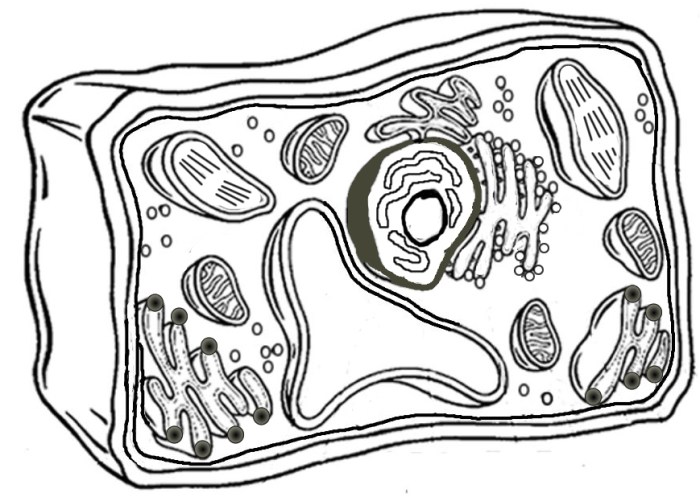
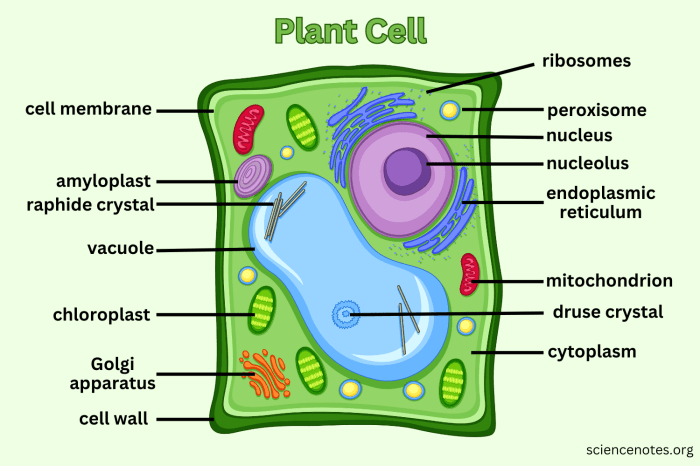
Plant cells share some similarities with animal cells, but possess several unique features. Like animal cells, they have a cell membrane, cytoplasm, nucleus, mitochondria, ribosomes, and a Golgi apparatus. However, plant cells are distinguished by the presence of a rigid cell wall, providing structural support and protection. A large central vacuole occupies a significant portion of the plant cell’s volume, storing water and other substances.
Furthermore, plant cells contain chloroplasts, the sites of photosynthesis, where light energy is converted into chemical energy.
Key Differences Between Animal and Plant Cells
The most striking differences between animal and plant cells are readily apparent in diagrams. Plant cells possess a rigid cell wall external to the cell membrane, a feature absent in animal cells. The large central vacuole is another defining characteristic of plant cells, typically absent or much smaller in animal cells. Finally, the presence of chloroplasts, responsible for photosynthesis, is exclusive to plant cells.
These three structures – cell wall, central vacuole, and chloroplasts – are easily distinguishable in comparative diagrams.
Textual Diagram of an Animal Cell
Imagine a circle (the cell membrane). Inside, a slightly smaller, irregularly shaped circle represents the nucleus. Scattered within the larger circle are numerous small dots (ribosomes), several oblong shapes (mitochondria), and a slightly curved, flattened sac-like structure (Golgi apparatus). Smaller, irregularly shaped vesicles (lysosomes) are also present throughout the cytoplasm.
Textual Diagram of a Plant Cell
Envision a rectangle (the cell wall) encompassing a circle (the cell membrane). Inside, a large, centrally located circle represents the central vacuole. A smaller, irregularly shaped circle within the cytoplasm is the nucleus. Scattered throughout the cytoplasm are small dots (ribosomes), oblong shapes (mitochondria), a slightly curved, flattened sac-like structure (Golgi apparatus), and numerous small, disc-shaped structures (chloroplasts).
Illustrative Examples

Let’s explore some visual representations of animal and plant cells, along with various coloring techniques and size comparisons to enhance understanding. These examples aim to provide a clear and engaging way to visualize the intricate structures within these fundamental units of life.
A Colored Animal Cell Diagram
Imagine a vibrant animal cell, its nucleus a rich, deep purple, centrally located and commanding attention. The surrounding cytoplasm is a soft, pale yellow, speckled with numerous smaller organelles. The rough endoplasmic reticulum (RER), appearing as a network of interconnected, flattened sacs, is colored a light teal, highlighting its role in protein synthesis. The smooth endoplasmic reticulum (SER), depicted as a network of interconnected tubules, is a contrasting light orange, reflecting its involvement in lipid metabolism.
The Golgi apparatus, a stack of flattened membranous sacs, is represented in a warm golden brown, suggesting its role in packaging and processing proteins. Mitochondria, the powerhouses of the cell, are shown as elongated, crimson-colored structures, their internal cristae subtly hinted at with darker shading. Lysosomes, small, spherical organelles, are a deep magenta, symbolizing their role in waste breakdown. Finally, the ribosomes, tiny dots scattered throughout the cytoplasm, are a muted grey-blue, their small size emphasizing their multitude.
A Colored Plant Cell Diagram
A plant cell, in contrast, presents a different visual spectacle. The large central vacuole dominates, taking up a significant portion of the cell’s volume and colored a calming, light seafoam green, signifying its role in turgor pressure and storage. The cell wall, the rigid outer boundary, is depicted in a sturdy, deep forest green, contrasting sharply with the vibrant contents within.
The chloroplasts, the sites of photosynthesis, are illustrated as numerous, oval-shaped structures, a bright, healthy lime green, their internal thylakoid membranes suggested by subtle darker shading. The nucleus, similar to the animal cell, is a deep purple, but positioned slightly off-center. The other organelles – mitochondria (crimson), endoplasmic reticulum (teal and orange), Golgi apparatus (golden brown), and ribosomes (muted grey-blue) – are present, but smaller and less prominent compared to the vacuole and chloroplasts.
Coloring Styles for Cell Diagrams
Several coloring styles can effectively illustrate cell diagrams. A realistic style might employ subtle shading and gradients to create a three-dimensional effect, mimicking the appearance of organelles under a microscope. A stylized approach, conversely, might use bold, flat colors and simplified shapes for a more graphic and easily digestible representation. Other options include using a cartoon style, emphasizing the fun and educational aspects, or employing a minimalist approach focusing on clear labeling and organizational clarity.
Example Color Palettes
For animal cells, a palette of deep purple (nucleus), pale yellow (cytoplasm), teal (RER), orange (SER), golden brown (Golgi), crimson (mitochondria), magenta (lysosomes), and muted grey-blue (ribosomes) creates a vibrant and informative image. For plant cells, a palette of light seafoam green (vacuole), deep forest green (cell wall), lime green (chloroplasts), deep purple (nucleus), and the same colors used for the animal cell organelles for consistency, produces a visually distinct representation.
Relative Sizes of Organelles
| Organelle | Animal Cell (Relative Size) | Plant Cell (Relative Size) |
|---|---|---|
| Nucleus | Large | Medium |
| Mitochondria | Small | Small |
| Ribosomes | Very Small | Very Small |
| Golgi Apparatus | Medium | Medium |
| Endoplasmic Reticulum | Large | Medium |
| Vacuole | Absent | Very Large |
| Cell Wall | Absent | Large |
| Chloroplasts | Absent | Medium |
Delving into the intricate artistry of animal and plant cell diagram coloring, one discovers a miniature world of vibrant hues and structural wonders. This microscopic beauty finds a kindred spirit in the majestic landscapes of the Arctic, where creatures of breathtaking design roam; for a delightful extension of this cellular exploration, consider the charming selection available at arctic animal coloring pages printable.
Returning to the cellular realm, the coloring of these diagrams offers a unique path to understanding the fundamental building blocks of life, both large and small.


